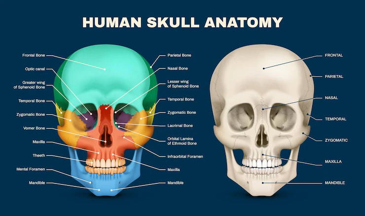
A skull diagram is one of the most useful tools for understanding the structure of the human head. It shows the different bones that make up the skull and helps students, teachers, medical professionals, and artists identify each part easily.
The human skull protects the brain, supports the face, and provides attachment points for muscles. Whether you are studying biology, anatomy, dentistry, nursing, or medicine, learning to read a skull diagram is an important skill.
This guide explains the human skull diagram, its parts, functions, and why it matters in education and healthcare.
What Is a Skull Diagram?
A skull diagram is a labeled illustration of the human skull showing its bones and important anatomical landmarks. It helps people understand how the skull is organized and how each bone contributes to protecting the brain and supporting facial structures.
Skull diagrams may show:
- Front view
- Side (lateral) view
- Top view
- Bottom view
- Internal view
- Labeled or unlabeled versions for study
Students commonly use labeled diagrams during anatomy classes and exam preparation.
Main Parts of the Human Skull
The adult human skull contains 22 bones, excluding the tiny ear bones.
These bones are divided into two major groups:
1. Cranial Bones
The cranial bones surround and protect the brain.
They include:
- Frontal bone
- Parietal bones (2)
- Temporal bones (2)
- Occipital bone
- Sphenoid bone
- Ethmoid bone
Together, these eight bones create the protective case around the brain.
2. Facial Bones
The facial bones form the structure of the face.
These include:
- Maxilla (2)
- Zygomatic bones (2)
- Nasal bones (2)
- Lacrimal bones (2)
- Palatine bones (2)
- Inferior nasal conchae (2)
- Vomer
- Mandible
The mandible is the only movable bone in the skull.
Functions of the Human Skull
The skull performs several important jobs.
Protects the Brain
Its primary role is shielding the brain from injury.
Supports the Face
The skull provides shape and structure for the eyes, nose, mouth, and cheeks.
Holds the Teeth
The upper jaw (maxilla) and lower jaw (mandible) support the teeth.
Enables Movement
The jaw allows chewing, speaking, and facial expressions.
Protects Sense Organs
The skull surrounds the:
- Eyes
- Ears
- Nose
These organs are protected by strong bony structures.
Important Bones in a Skull Diagram
Frontal Bone
Located at the forehead.
Functions:
- Forms the forehead
- Protects the frontal lobe
- Forms part of the eye sockets
Parietal Bones
These two bones make up the roof and sides of the skull.
Functions:
- Protect the upper brain
- Connect with neighboring bones through sutures
Temporal Bones
Found on each side of the head.
Functions:
- House the hearing organs
- Support jaw movement
- Protect parts of the brain
Occipital Bone
Located at the back of the skull.
Functions:
- Protects the back of the brain
- Contains the opening where the spinal cord enters the skull
Sphenoid Bone
Often called the “keystone” bone because it connects many skull bones.
Functions:
- Supports the skull base
- Forms part of the eye socket
Ethmoid Bone
Located between the eyes.
Functions:
- Supports the nasal cavity
- Forms part of the eye sockets
Mandible
The lower jawbone.
Functions:
- Chewing
- Speaking
- Facial movement
It is attached to the skull through the temporomandibular joint (TMJ).
Skull Sutures
Sutures are fibrous joints connecting skull bones.
Major sutures include:
- Coronal suture
- Sagittal suture
- Lambdoid suture
- Squamous suture
These joints are fixed in adults but remain flexible in infants.
Openings Found in the Skull
Several important holes allow nerves and blood vessels to pass.
Examples include:
- Foramen magnum
- Optic canal
- Jugular foramen
- Infraorbital foramen
These openings are commonly labeled in advanced skull diagrams.
Skull Diagram Views
Different views highlight different anatomical features.
Front View
Shows:
- Eye sockets
- Nasal cavity
- Maxilla
- Mandible
Side View
Displays:
- Temporal bone
- Zygomatic arch
- Mandible
- Occipital bone
Superior View
Shows:
- Frontal bone
- Parietal bones
- Sagittal suture
Inferior View
Highlights:
- Hard palate
- Foramen magnum
- Occipital condyles
Why Students Use Skull Diagrams
A skull diagram makes anatomy easier to understand.
Students use it for:
- Biology classes
- Medical school
- Dental education
- Nursing programs
- Physical therapy
- Radiology
- Exam preparation
Visual learning improves memory and makes identifying bones much easier.
Clinical Importance of Skull Anatomy
Understanding skull anatomy helps healthcare professionals diagnose and treat many conditions.
Examples include:
Skull Fractures
Head injuries may fracture one or more skull bones.
TMJ Disorders
Problems involving the jaw joint can affect speaking and chewing.
Congenital Conditions
Some babies are born with abnormal skull development, such as craniosynostosis.
Brain Surgery
Neurosurgeons rely on detailed knowledge of skull anatomy before performing procedures.
Dental Treatments
Dentists study the skull to understand jaw alignment and tooth positioning.
Tips for Memorizing a Skull Diagram
Learning skull anatomy becomes easier with practice.
Helpful strategies include:
- Study labeled diagrams first.
- Learn bones in groups.
- Use flashcards.
- Practice drawing the skull.
- Test yourself with unlabeled diagrams.
- Review anatomy regularly.
Many students find color-coded diagrams especially useful.
Common Mistakes When Studying Skull Anatomy
Avoid these common errors:
- Confusing the maxilla with the mandible.
- Forgetting that the mandible is movable.
- Mixing up cranial and facial bones.
- Ignoring skull sutures.
- Memorizing names without understanding functions.
Understanding both structure and function improves long-term learning.
Interesting Facts About the Human Skull
- Adults have 22 skull bones.
- Babies are born with soft spots called fontanelles.
- The skull reaches most of its adult size during childhood.
- The mandible is the strongest facial bone.
- Skull thickness varies in different areas.
Conclusion
A skull diagram is an essential learning tool for understanding human anatomy. It clearly illustrates the bones, sutures, and landmarks that protect the brain and support the face. Whether you’re a student, healthcare professional, or simply curious about the human body, studying a skull diagram helps build a solid foundation in anatomy.
By learning the names, locations, and functions of each bone, you can better understand how the skull supports everyday activities like speaking, chewing, seeing, and protecting the brain.
Frequently Asked Questions (FAQs)
1. What is a skull diagram?
A skull diagram is a labeled illustration showing the bones and anatomical structures of the human skull for educational and medical purposes.
2. How many bones are in the human skull?
The adult human skull contains 22 bones: 8 cranial bones and 14 facial bones.
3. Which bone is movable in the skull?
The mandible (lower jaw) is the only movable bone in the human skull.
4. Why is a skull diagram important?
It helps students and healthcare professionals learn bone names, locations, functions, and anatomical relationships.
5. What are skull sutures?
Skull sutures are immovable fibrous joints that connect the bones of the skull.
6. What is the main function of the skull?
The skull protects the brain, supports the face, houses the sense organs, and provides attachment points for muscles.
7. Who commonly uses skull diagrams?
Medical students, biology students, dentists, nurses, doctors, physical therapists, and anatomy instructors frequently use skull diagrams.
8. Can I learn skull anatomy using diagrams alone?
Diagrams are an excellent starting point, but combining them with textbooks, 3D models, and practical study provides a deeper understanding of skull anatomy.